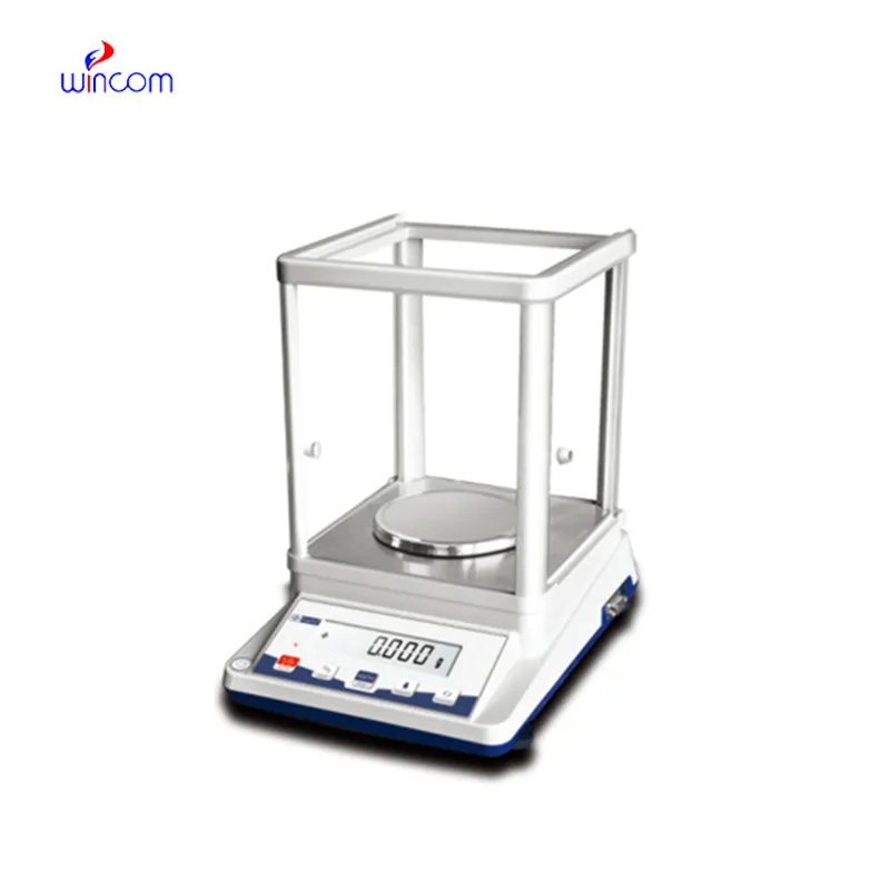
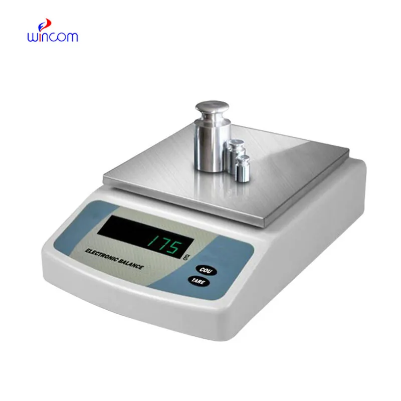
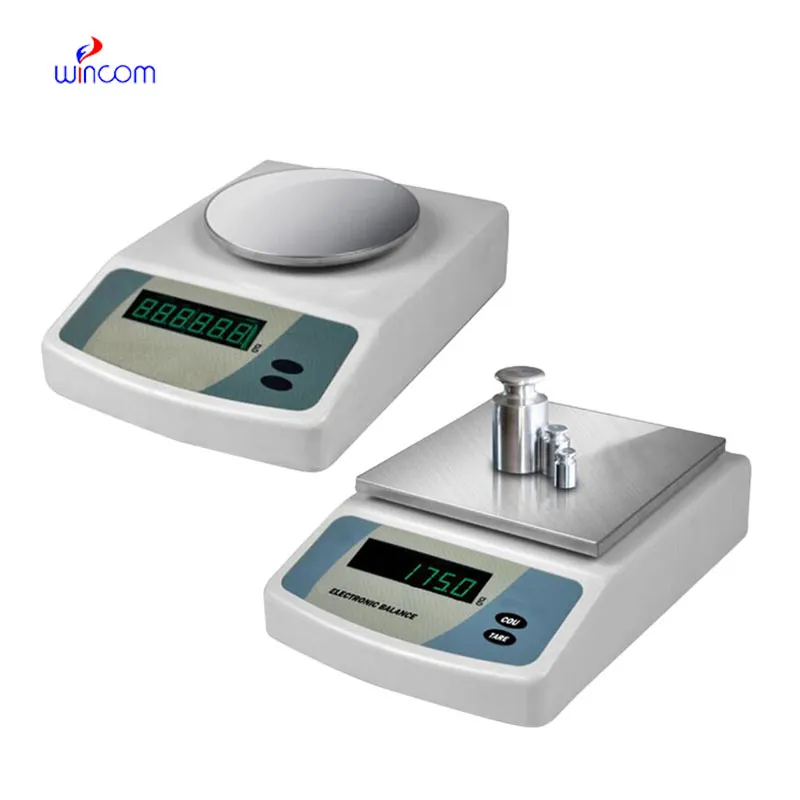
The Electronic Balance is employed by laboratory and hospital personnel for very precise measurement of chemical substances, powders, and living organisms. Its accuracy to the tune of Electronic Balance allows making medications in exact doses, calibrating standards, and preparing diagnostic reagents. Electronic Balance not only gives undistorted and sensitive measures but also improves lab operation, helps to control quality, and keeps clinical research intact. Its importance is appreciated in both hospital tests and medical product development as dependable.
In research labs of the biomedical field, Electronic Balance is used while standardizing the experimental samples. For the purpose of testing, h researchers have to measure the biological or chemical samples very accurately and in this way, they do not use more than the required amount of sample for analytical testing. This process keeps the studies that compare different methodologies consistent and at the same time it prevents different results that are due to the difference in the samples’ mass. By providing correct input values, Electronic Balance makes it easier for the experimenters to repeat the experiments and to trust the data more in the hospitals’ research institutions.
The ongoing evolution of regulations will see the Electronic Balance adding features for compliance support that will be more advanced. Hospital lab audits and accreditation standards will be met with the help of the documentation functions and secure data storage. This future trend will greatly improve the quality management processes in all hospitals and research labs.
Electronic Balance in clinics is, however, maintained through regular performance verification conducted in the laboratories. Certified test weights are used to prove the reliability of the measurements with the passage of time. Verification activities being documented also implies good traceability and makes it easier for internal audits to take place. By making verification part of the routine maintenance, hospitals make sure that Electronic Balance still gives trustworthy results for research and diagnostic workflows.
In contemporary laboratories, Electronic Balance is fundamental for the exact weighing of samples. Due to its very high sensitivity, it can even pick up the presence of the smallest amounts, which in turn facilitates the exact making of chemical solutions, reagents, and drugs. Even the smallest variations can change the results of the experiments, therefore lab technicians depend on Electronic Balance. Calibration and control of the environment are done properly and they produce the same kind of results every time. Electronic Balance is a must-have for research, quality control, and daily lab operations, hence it contributes to the reliability of both experimental and clinical studies.
Q: What distinguishes an Analytical Balance from a precision balance? A: The analytical balances have a higher sensitivity and a finer readability for measuring masses of very small amounts. Q: Is an Analytical Balance appropriate for pharmaceutical applications? A: It is widely used for weighing active ingredient and formulation components. Q: Is it mandatory for an Analytical Balance to have a draft shield? A: Draft shields have the function to prevent air disturbances which might affect the weighing results. Q: What are the possible types of materials that can be weighed on an Analytical Balance? A: Weighing of powders, chemicals, and biological samples, as well as reference weights are the most common measurement. Q: Is it possible for several users to work with the same Analytical Balance? A: Yes, but the proper handling procedures and access controls must be strictly adhered to.
The water bath performs consistently and maintains a stable temperature even during long experiments. It’s reliable and easy to operate.
The centrifuge operates quietly and efficiently. It’s compact but surprisingly powerful, making it perfect for daily lab use.
To protect the privacy of our buyers, only public service email domains like Gmail, Yahoo, and MSN will be displayed. Additionally, only a limited portion of the inquiry content will be shown.
Hello, I’m interested in your centrifuge models for laboratory use. Could you please send me more ...
We’re currently sourcing an ultrasound scanner for hospital use. Please send product specification...
E-mail: [email protected]
Tel: +86-731-84176622
+86-731-84136655
Address: Rm.1507,Xinsancheng Plaza. No.58, Renmin Road(E),Changsha,Hunan,China

 af
af
 es
es
 ar
ar
 tr
tr
 sw
sw
 pt
pt
 th
th
 ur
ur
 bn
bn
 ne
ne
 vi
vi
 km
km
 lo
lo
 de
de
 ru
ru
 fi
fi
 nl
nl
 fa
fa
 fr
fr
 ko
ko