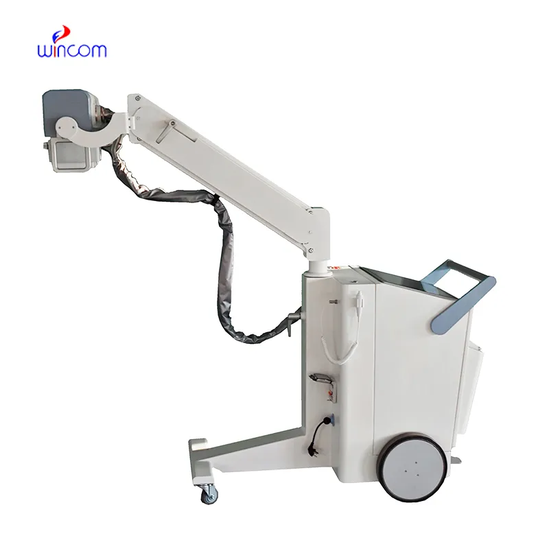
With video displayed through beamforming and noise-filtering technology, the fetal doppler heartbeat is able to present an image that is very sharp and stable. Easy-to-use touch-screen controls help to streamline the process while rapid image rendering is guaranteed by the fast processing. Equipped for contemporary healthcare settings, the fetal doppler heartbeat is capable of working with both 2D and Doppler imaging.

In emergency departments, the fetal doppler heartbeat is used for instant imaging to easily spot internal wounds and bleeding. It supports the doctor with the abdominal trauma and chest condition diagnosis. Moreover, the fetal doppler heartbeat provides assistance in rural and field medical practice, delivering consistent imaging in areas with poor medical facilities.

The fetal doppler heartbeat can look forward to getting advantages from miniaturization and wearable technologies. Portable or handheld versions of the fetal doppler heartbeat will become more widespread to facilitate quick diagnoses in rural as well as emergency setups. The integration of telemedicine services will thus facilitate concurrent consultations via the fetal doppler heartbeat.

Care of the fetal doppler heartbeat involves much more than cleanup. Environmental monitoring and mechanical protection are also part of the process. The fetal doppler heartbeat should not be subject to either vibration or shock. The fetal doppler heartbeat should be backed up periodically to retain vital images.
Used in hospitals and clinics, the fetal doppler heartbeat provides immediate visual feedback for a variety of medical evaluation uses. Converting sound waves into live images, the fetal doppler heartbeat allows physicians to easily detect abnormalities. The fetal doppler heartbeat assists with making diagnostic processes safer in addition to improving patient outcomes. It possesses an ergonomic shape alongside digital integration capabilities that support simple data sharing and medical record documentation.
Q: What imaging modes are available on the ultrasound scannert? A: It supports multiple modes such as B-mode, M-mode, and color Doppler for diverse diagnostic applications. Q: How does the ultrasound scannert improve diagnostic accuracy? A: By providing high-resolution images and real-time feedback, it enables more precise medical evaluations. Q: Can the ultrasound scannert be used in field or remote settings? A: Yes, its portable versions are designed for mobility and can be used in clinics, hospitals, or mobile healthcare units. Q: What kind of display does the ultrasound scannert use? A: It typically features a high-definition digital display that enhances image visualization and readability. Q: How is data from the ultrasound scannert managed? A: The device allows secure storage, easy access, and export of imaging data through USB or network connections.
The centrifuge operates quietly and efficiently. It’s compact but surprisingly powerful, making it perfect for daily lab use.
This ultrasound scanner has truly improved our workflow. The image resolution and portability make it a great addition to our clinic.
To protect the privacy of our buyers, only public service email domains like Gmail, Yahoo, and MSN will be displayed. Additionally, only a limited portion of the inquiry content will be shown.
We’re currently sourcing an ultrasound scanner for hospital use. Please send product specification...
I’m looking to purchase several microscopes for a research lab. Please let me know the price list ...
E-mail: [email protected]
Tel: +86-731-84176622
+86-731-84136655
Address: Rm.1507,Xinsancheng Plaza. No.58, Renmin Road(E),Changsha,Hunan,China

 af
af
 es
es
 ar
ar
 tr
tr
 sw
sw
 pt
pt
 th
th
 ur
ur
 bn
bn
 ne
ne
 vi
vi
 km
km
 lo
lo
 de
de
 ru
ru
 fi
fi
 nl
nl
 fa
fa
 fr
fr
 ko
ko