Through a combination of novel imaging software and the fetal heart sound by doppler, the professionals can be very precise with the details of the anatomy they have captured and even more so with the accuracy of the final result. An ergonomic design of the device contributes to the reduction of operator fatigue during long use. The device, the fetal heart sound by doppler, also comes with the ability to connect to more than one probe thus giving it the flexibility required when catering to various diagnostic needs. Besides that, the data export and connection functions of the device help to decrease the time taken in report-generating which is done through image sharing.

In medical imaging, the fetal heart sound by doppler is a trustworthy device for different clinical departments. It provides vascular studies support by observing the state of the arteries and veins; in urology, it has a role in the assessment of the bladder and prostate health. The fetal heart sound by doppler also enables emergency medical staff to make quick trauma evaluations, directing their actions precisely and efficiently.

The next-generation fetal heart sound by doppler solutions come with better processing capabilities and intelligent algorithms that improve the clarity of images in addition to lessening reliance on operators. The aspect of augmented reality will change the world of surgical operations. The fetal heart sound by doppler solutions will also change the face of delivering healthcare by facilitating quicker and more accurate diagnoses.

For long-term functionality, it is recommended that the fetal heart sound by doppler remain within an environment that maintains controlled levels of both humidity and temperature. The cables should be unwound slowly to ensure that no undue stress or wire breakages occur. The fetal heart sound by doppler should also be properly disinfected each time a patient has been examined.
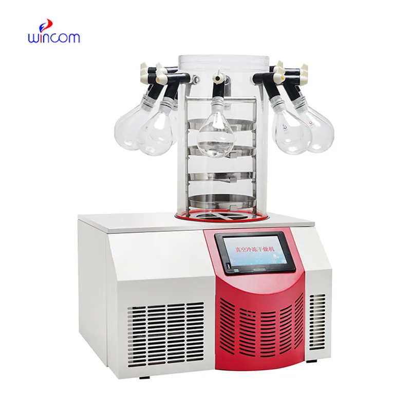
The fetal heart sound by doppler is an essential diagnostic modality in the modern healthcare system, permitting the non-invasive imaging of internal organs and tissues. By transmitting sound waves and reading their echoes, it provides real-time data on physiology. The fetal heart sound by doppler makes precise diagnoses feasible in all specialties, improving clinical decision-making and patient confidence.
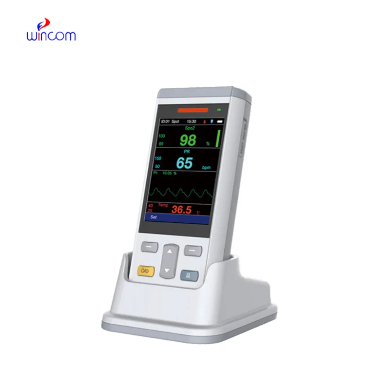
Q: What makes the ultrasound scannert effective for diagnostic imaging? A: Its high-frequency sound wave technology allows accurate visualization of internal body structures in real time. Q: How portable is the ultrasound scannert? A: The device features a compact and lightweight design, allowing easy movement between clinical departments. Q: What types of probes are compatible with the ultrasound scannert? A: It supports multiple probe types, including linear, convex, and phased array probes for varied diagnostic needs. Q: Does the ultrasound scannert require special training to operate? A: Basic technical training is recommended to maximize its imaging performance and functionality. Q: How long can the ultrasound scannert operate continuously? A: It is designed for extended use with efficient cooling systems and stable power performance.
We’ve used this centrifuge for several months now, and it has performed consistently well. The speed control and balance are excellent.
This ultrasound scanner has truly improved our workflow. The image resolution and portability make it a great addition to our clinic.
To protect the privacy of our buyers, only public service email domains like Gmail, Yahoo, and MSN will be displayed. Additionally, only a limited portion of the inquiry content will be shown.
We’re looking for a reliable centrifuge for clinical testing. Can you share the technical specific...
We’re interested in your delivery bed for our maternity department. Please send detailed specifica...
E-mail: [email protected]
Tel: +86-731-84176622
+86-731-84136655
Address: Rm.1507,Xinsancheng Plaza. No.58, Renmin Road(E),Changsha,Hunan,China

 af
af
 es
es
 ar
ar
 tr
tr
 sw
sw
 pt
pt
 th
th
 ur
ur
 bn
bn
 ne
ne
 vi
vi
 km
km
 lo
lo
 de
de
 ru
ru
 fi
fi
 nl
nl
 fa
fa
 fr
fr
 ko
ko