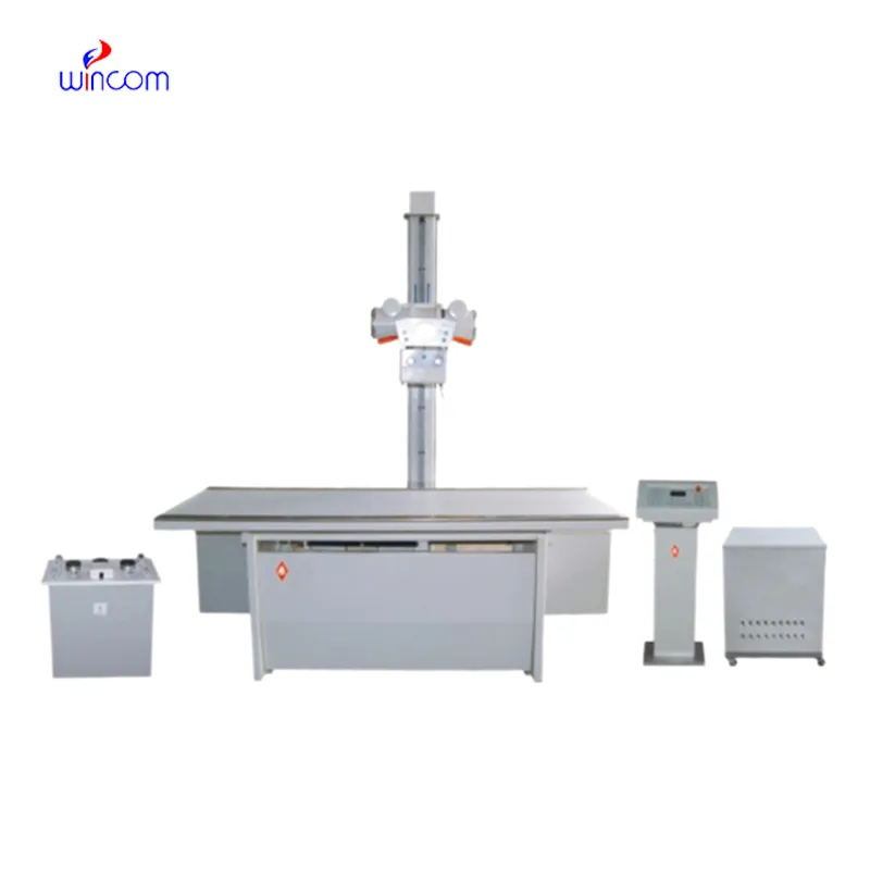
The invention of the x ray machine comes equipped with advanced digital detectors that transform X-ray energy into high definition images of incredible detail. The system's design makes it easy to use and facilitates quick image capturing. The invention of the x ray machine system can be connected effortlessly to hospital information systems that enable the secure transfer of data. The system's robust design provides support for long-term use within healthcare settings.

The invention of the x ray machine is used in numerous various clinical departments, including orthopedics, radiology, and oncology. It is usually applied to detect lung lesions and bone fractures as well as track tumor growth. In children's practice, the invention of the x ray machine enables safe imaging with child-friendly doses suitable for diagnostic needs in children.
The invention of the x ray machine of the future will target integrating artificial intelligence to aid image interpretation and identify anomalies. Analysis software will automatically detect early-stage diseases more accurately. The invention of the x ray machine will further feature low-dose radiation technologies, which will ensure that imaging is safer, more sustainable for both patients and operators.
Care and maintenance of the invention of the x ray machine are required to ensure repeat imaging quality and ruggedness. Cable, detector, and collimator faults are averted by periodic checks. The invention of the x ray machine need to be kept in a dust-free environment with low temperatures to avoid overheating and dust depositing on them. Routine calibration and radiation output monitor checks ensure accurate diagnostic data.
The invention of the x ray machine turns X-ray radiation into visual information that offers clarity on skeletal and soft tissues. The digital imaging technology of the invention of the x ray machine helps improve the clarity of images by reducing radiation. The invention of the x ray machine helps healthcare providers evaluate a patient's skeletal system since it assists in diagnosing fractures, lung disease, and dental problems.
Q: What makes an x-ray machine different from a CT scanner? A: An x-ray machine captures a single 2D image, while a CT scanner takes multiple x-rays from different angles to create 3D cross-sectional views. Q: How is image quality measured in an x-ray machine? A: Image quality depends on factors like contrast, resolution, and exposure settings, which are adjusted based on the target area being examined. Q: What power supply does an x-ray machine require? A: Most x-ray machines operate on high-voltage power systems, typically between 40 to 150 kilovolts, depending on their intended use. Q: Can x-ray machines be used for dental imaging? A: Yes, specialized dental x-ray machines provide detailed images of teeth, jaws, and surrounding structures to support oral health assessments. Q: How does digital imaging improve x-ray efficiency? A: Digital systems allow instant image preview, faster diagnosis, and reduced need for retakes, improving workflow efficiency in clinical environments.
We’ve used this centrifuge for several months now, and it has performed consistently well. The speed control and balance are excellent.
We’ve been using this mri machine for several months, and the image clarity is excellent. It’s reliable and easy for our team to operate.
To protect the privacy of our buyers, only public service email domains like Gmail, Yahoo, and MSN will be displayed. Additionally, only a limited portion of the inquiry content will be shown.
Could you please provide more information about your microscope range? I’d like to know the magnif...
I’m looking to purchase several microscopes for a research lab. Please let me know the price list ...
E-mail: [email protected]
Tel: +86-731-84176622
+86-731-84136655
Address: Rm.1507,Xinsancheng Plaza. No.58, Renmin Road(E),Changsha,Hunan,China

 af
af
 es
es
 ar
ar
 tr
tr
 sw
sw
 pt
pt
 th
th
 ur
ur
 bn
bn
 ne
ne
 vi
vi
 km
km
 lo
lo
 de
de
 ru
ru
 fi
fi
 nl
nl
 fa
fa
 fr
fr
 ko
ko