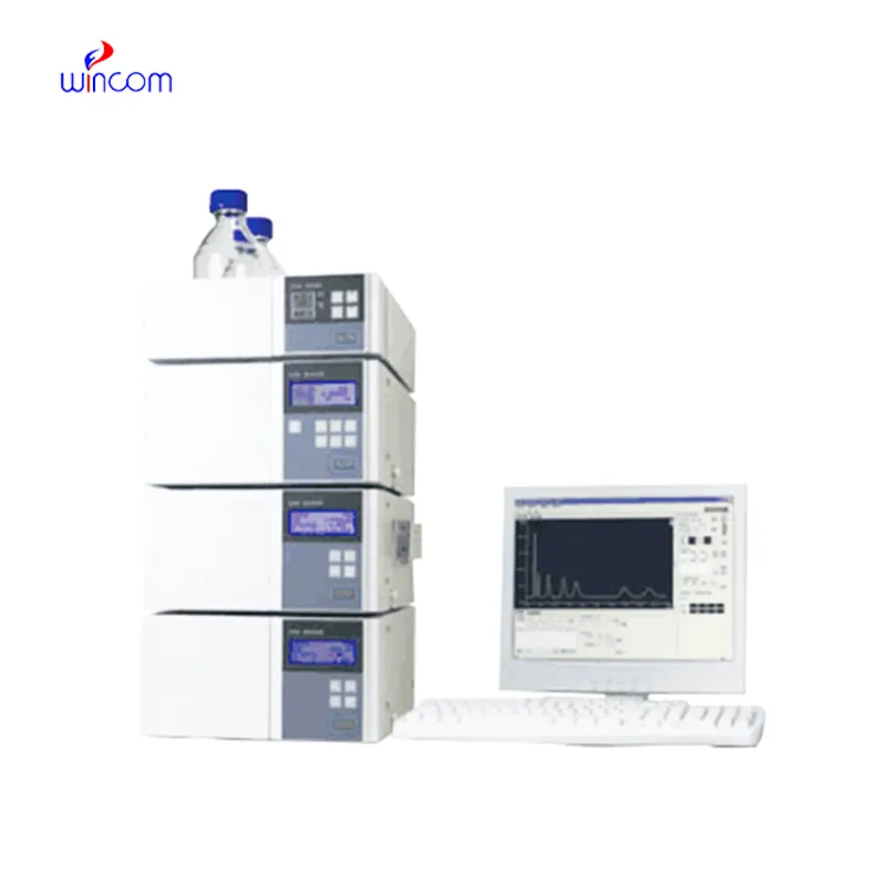
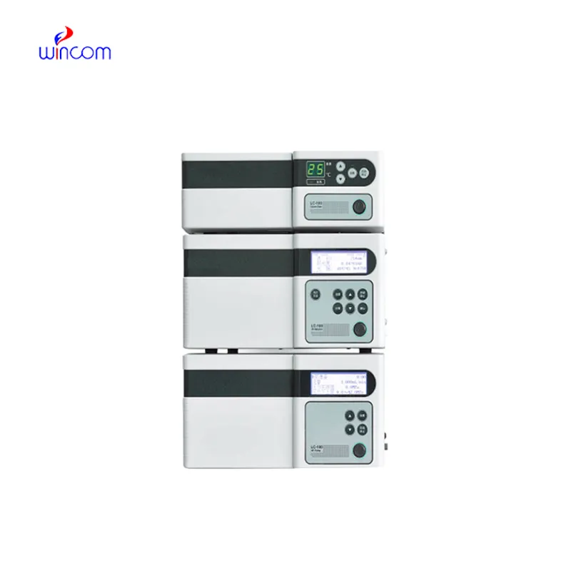
In medical and clinical laboratories, the use of ion exchange hplc results in highly precise determination of therapeutic compounds, metabolites, and biochemical markers. It facilitates creation of detailed patient sample profiles for research and diagnostics. The laboratory personnel prefer ion exchange hplc for confirming method reproducibility, validating analytical procedures, and keeping track of sample integrity. The ultrahigh sensitivity and versatility of the apparatus permit the laboratories to cater to varied applications, thus helping hospitals and research centers to provide reliable and accurate analytical results in various fields of science.

ion exchange hplc finds use in clinical toxicology laboratories to pinpoint and measure the amounts of possible poisons or drugs in abuse samples taken from patients. It is based on the separation of the various substances from complex mixtures like blood or urine, and that information is very important for the hospital doctors, who will then diagnose the case, decide on the treatment and monitor the patient’s safety.
The instruments for ion exchange hplc of the future will be equipped with separation methods in multiple dimensions and fully automated sample preparation. The detection of trace amounts of metabolites, drugs, and biomarkers will be so accurate that hospitals and clinical laboratories will be the first to reap the benefits. The applications of ion exchange hplc in the future will greatly help in complex diagnostics, research studies, and laboratory efficiency.
Regular system checks, cleaning of detector flow cells, and changing consumable parts whenever necessary are some of the actions that the laboratory staff should take in order to keep the ion exchange hplc working efficiently. Observing pump performance, taking care of solvent contamination, and storing columns correctly prolong the life of the instrument. Good maintenance assures reproducibility, cuts down on time without access to equipment, and promotes high-quality analysis in hospitals and clinical labs.
ion exchange hplc is of utmost importance in biochemistry laboratories of both universities and hospitals. It makes detailed study of proteins, peptides, and metabolites possible through the separation of intricate mixtures. The application of it includes but is not limited to enzymatic analysis, biomarker detection, and data obtained through metabolomics. The sensitivity and reproducibility of the device guarantee genuine molecular profiles. Lab technicians make use of ion exchange hplc to conclude their experiments and provide evidence for scientific publications. Its accuracy and versatility give biochemistry labs the ability to perform cutting-edge research in molecular mechanisms, disease pathways, and therapy targets thus, it becomes an indispensable tool for both analytical and clinical lab investigations.
Q: Do you need special training for HPLC operation? A: The answer is yes, training is a prerequisite to accurately and safely using pumps, columns, and detectors. Q: What type of maintenance does HPLC have? A: It requires cleaning, flushing, and inspection of all components as well as calibrating. Q: Is it possible to use HPLC in drug monitoring? A: Sure, it is a common practice in hospitals to monitor the levels of therapeutic drugs and also to identify metabolites in the samples taken from the patients. Q: What is the duration of analysis using HPLC in a typical case? A: The analysis time can range from a few minutes to more than an hour depending on the nature of the sample and the kind of column used. Q: Is HPLC a good choice for environmental testing? A: Yes, it can be used to find out the presence of pollutants, pesticides, and other harmful substances in water, soil, and air samples.
The delivery bed is well-designed and reliable. Our staff finds it simple to operate, and patients feel comfortable using it.
We’ve been using this mri machine for several months, and the image clarity is excellent. It’s reliable and easy for our team to operate.
To protect the privacy of our buyers, only public service email domains like Gmail, Yahoo, and MSN will be displayed. Additionally, only a limited portion of the inquiry content will be shown.
Could you please provide more information about your microscope range? I’d like to know the magnif...
We’re looking for a reliable centrifuge for clinical testing. Can you share the technical specific...
E-mail: [email protected]
Tel: +86-731-84176622
+86-731-84136655
Address: Rm.1507,Xinsancheng Plaza. No.58, Renmin Road(E),Changsha,Hunan,China

 af
af
 es
es
 ar
ar
 tr
tr
 sw
sw
 pt
pt
 th
th
 ur
ur
 bn
bn
 ne
ne
 vi
vi
 km
km
 lo
lo
 de
de
 ru
ru
 fi
fi
 nl
nl
 fa
fa
 fr
fr
 ko
ko