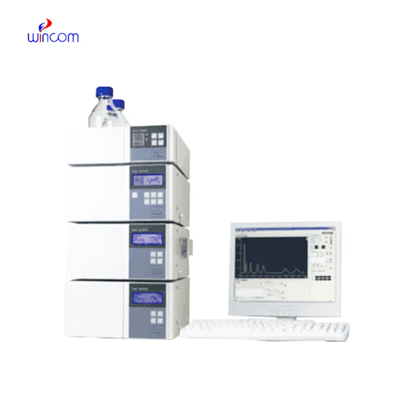
preparative hplc column is a critical technique to obtain analytical information in studies of medicines, clinical samples, and biochemistry. It isolates compounds according to their chemical characteristics, generating reproducible analytical results. Laboratory scientists use preparative hplc column to perform drug stability tests, monitor patient biomarkers, and find impurities. Its very high accuracy and flexibility allow thorough sample analysis in research, hospital, and clinical laboratory environments, thus becoming a fundamental device for assuring precision in both experimental and diagnostic results.

Hospital laboratories depend on preparative hplc column for identifying minute quantities of pharmaceuticals and therapeutic agents in difficult-to-analyze biological samples. Its use spans drug compliance testing, pharmacokinetics profiling, and tracking medications after surgery. The laboratory personnel can rely on it for exact measurement, thus increasing the efficiency of clinical treatment.
The forthcoming breed of preparative hplc column will put a spotlight on intelligent instruments that are connected with cloud-based surveillance. Through this monitoring, hospitals will be able to gain a remote view of laboratory activities and the results of sample analysis. Lab productivity will be greatly increased by the upcoming preparative hplc column, and together with the new features, patient testing and therapy monitoring even in difficult clinical settings will be more accurate.
Routine upkeep of preparative hplc column is of utmost importance in clinical laboratories to maintain the accuracy of patient sample analysis. Regular cleaning of pipes, changing of deteriorated seals and calibration of measuring instruments will block adulteration and keep the latter's sensitivity. Lab personnel must record maintenance activities and keep watch over system performance. Constant attention guarantees that preparative hplc column provides dependable, reproducible results for hospital diagnosis and research work.
Therapeutic drug monitoring relies heavily on preparative hplc column in hospital settings. It determines the concentration of drugs in the body to guarantee efficiency and security. The laboratory staff uses it for the examination of blood, serum, or urine samples, and signifies small molecular compounds with high accuracy. By yielding consistent outcomes, preparative hplc column services the medics in changing the amounts and preventing side effects. Its use goes to hormone level testing, metabolite analysis, and pharmacokinetics research. With quick processing and accurate information, preparative hplc column is a part of the hospital patient care, making evidence-based treatment decisions possible and enhancing clinical outcomes in different departments.
Q: What types of HPLC columns are available? A: Reversed-phase, normal-phase, ion-exchange, and size-exclusion columns are the main types of columns used according to the nature of the analytes. Q: Can multiple samples be analyzed simultaneously? A: Yes, in high-throughput systems, automated sample injection and sequential analysis are among the techniques to achieve this. Q: How does temperature affect HPLC performance? A: Temperature changes can cause variations in separation efficiency and retention times; however, the majority of labs make use of precise temperature control. Q: Can HPLC be integrated with data software? A: Sure, it can be linked with laboratory software for data collection, processing, and reporting. Q: What types of laboratories use HPLC? A: HPLC is employed by hospitals, pharmaceuticals, biochemistry research, and environmental testing labs.
We’ve used this centrifuge for several months now, and it has performed consistently well. The speed control and balance are excellent.
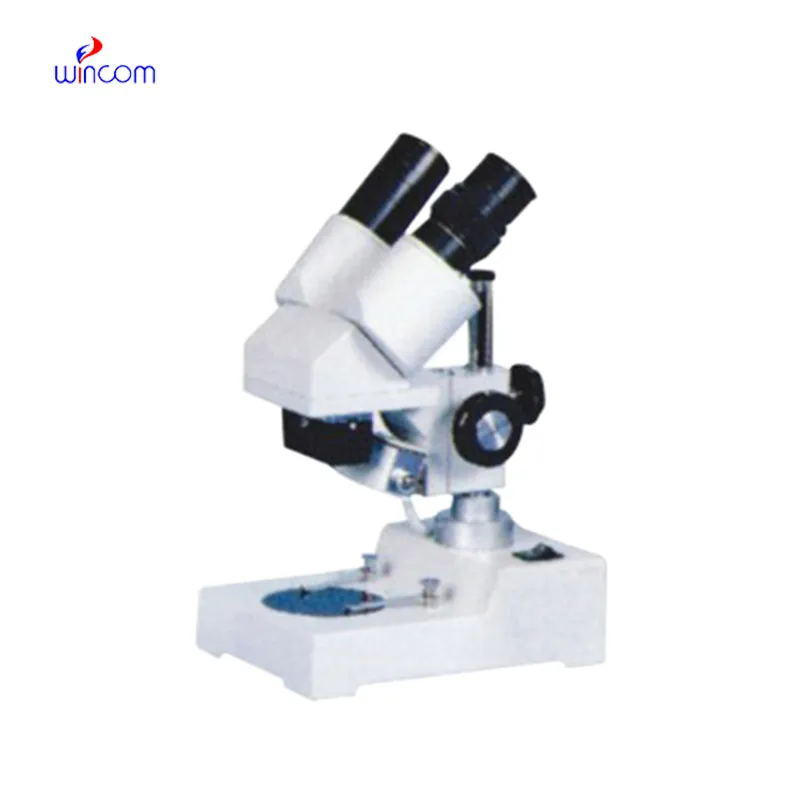
I’ve used several microscopes before, but this one stands out for its sturdy design and smooth magnification control.
To protect the privacy of our buyers, only public service email domains like Gmail, Yahoo, and MSN will be displayed. Additionally, only a limited portion of the inquiry content will be shown.
We are planning to upgrade our imaging department and would like more information on your mri machin...
Hello, I’m interested in your centrifuge models for laboratory use. Could you please send me more ...
E-mail: [email protected]
Tel: +86-731-84176622
+86-731-84136655
Address: Rm.1507,Xinsancheng Plaza. No.58, Renmin Road(E),Changsha,Hunan,China

 af
af
 es
es
 ar
ar
 tr
tr
 sw
sw
 pt
pt
 th
th
 ur
ur
 bn
bn
 ne
ne
 vi
vi
 km
km
 lo
lo
 de
de
 ru
ru
 fi
fi
 nl
nl
 fa
fa
 fr
fr
 ko
ko