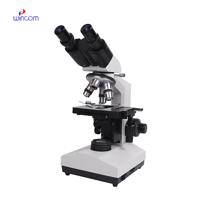
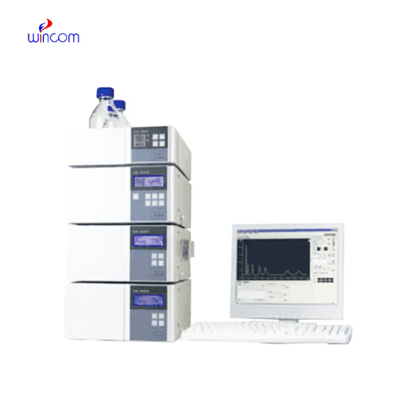
rp-hplc offers high resolution separation of complex samples in clinical, pharmaceutical, and hospital laboratories, thereby supporting advanced laboratory workflows. It allows performing an in-depth analysis of drugs, metabolites, and small biomolecules. rp-hplc is used by laboratory staff for research validation, patient monitoring, and method development. Its precision, speed, and adaptability make analytical efficiency greater and at the same time, make consistent and reproducible results which in turn, strengthen laboratory operations in the areas of healthcare and scientific environments.

Hospital laboratories depend on rp-hplc for identifying minute quantities of pharmaceuticals and therapeutic agents in difficult-to-analyze biological samples. Its use spans drug compliance testing, pharmacokinetics profiling, and tracking medications after surgery. The laboratory personnel can rely on it for exact measurement, thus increasing the efficiency of clinical treatment.
rp-hplc is assigned to become an important player in translational research which is being conducted in hospitals. Among the future developments are the combined detection systems, quicker analysis cycles, and improved reproducibility. rp-hplc will be the mainstay of hospitals' molecular profiling and drug testing along with patient monitoring thus facilitating hospital diagnostics and personalized medicine research.
Systematic cleaning, pressure monitoring, and timely worn parts replacement are among the measures to be taken in the hospital laboratories to keep rp-hplc under control. Laboratory staff must ensure the observance of the suggested operating conditions, avoid the formation of air bubbles in the system, and check for proper solvent compatibility. Regular maintenance maintains the performance of the column, avoids contamination, and allows the analysis to be precise and reproducible, thereby benefiting not only routine patient testing but also experimental research.
Clinical laboratories make use of rp-hplc to analyze patient samples with remarkable accuracy. It identifies biomarkers, metabolites, and the levels of therapeutic drugs, thus giving reliable information about the disease status and monitoring treatment. Sensitivity of the technique permits determination of compounds in very minute amounts, which is critical in clinical testing. By resolving complex composition, rp-hplc guarantees accurate and reproducible results for laboratory diagnostics. Lab staff utilizes it for daily testing, quality control, and research activities, thus making rp-hplc a vital part of contemporary clinical laboratory work that caters to patient care, treatment choices, and lab data integrity.
Q: What is the sample preparation for HPLC? A: For the most part, samples should be filtered, diluted, or subjected to solvent extraction in order to avoid column clogs and have the results be accurate Q: Is HPLC able to pick trace-level compounds? A: With the right detectors, it can pick up such substances in extremely small amounts with high sensitivity. Q: Is HPLC a method that can be applied to analysis of proteins? A: Yes, particularly if one employs size-exclusion and reversed-phase columns for protein, peptide, and biomolecule separation. Q: What is the process of calibrating HPLC? A: The process is done by taking standards of known concentrations that are the same as the one in the sample and using them to check the performance of the column and the accuracy of the detector. Q: Are particular solvents needed for HPLC? A: Yes, the solvents used need to be compatible with the type of the column and the detectors to prevent any damage or interference in the analysis process.
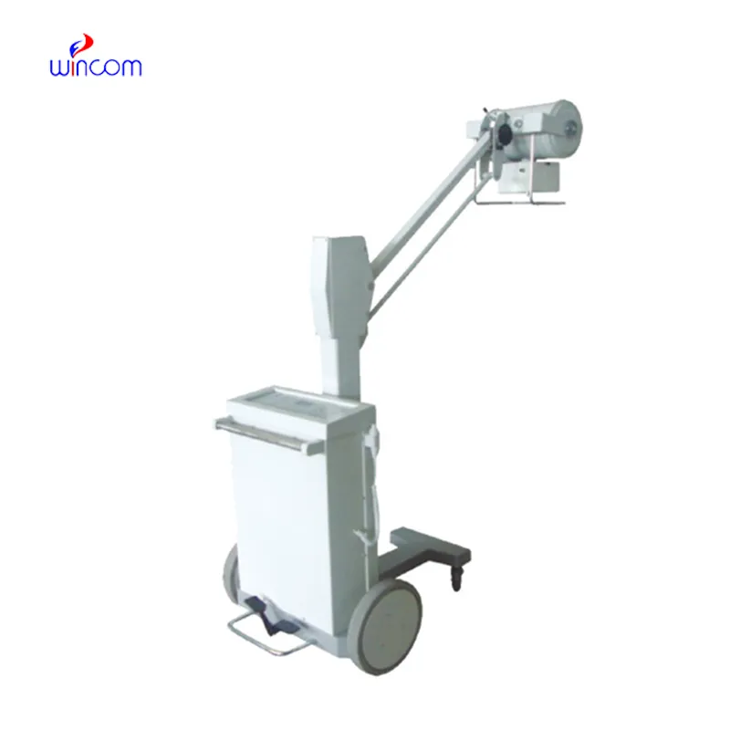
This x-ray machine is reliable and easy to operate. Our technicians appreciate how quickly it processes scans, saving valuable time during busy patient hours.
We’ve been using this mri machine for several months, and the image clarity is excellent. It’s reliable and easy for our team to operate.
To protect the privacy of our buyers, only public service email domains like Gmail, Yahoo, and MSN will be displayed. Additionally, only a limited portion of the inquiry content will be shown.
Hello, I’m interested in your water bath for laboratory applications. Can you confirm the temperat...
Could you share the specifications and price for your hospital bed models? We’re looking for adjus...
E-mail: [email protected]
Tel: +86-731-84176622
+86-731-84136655
Address: Rm.1507,Xinsancheng Plaza. No.58, Renmin Road(E),Changsha,Hunan,China

 af
af
 es
es
 ar
ar
 tr
tr
 sw
sw
 pt
pt
 th
th
 ur
ur
 bn
bn
 ne
ne
 vi
vi
 km
km
 lo
lo
 de
de
 ru
ru
 fi
fi
 nl
nl
 fa
fa
 fr
fr
 ko
ko