In medical and clinical laboratories, the use of size exclusion hplc columns results in highly precise determination of therapeutic compounds, metabolites, and biochemical markers. It facilitates creation of detailed patient sample profiles for research and diagnostics. The laboratory personnel prefer size exclusion hplc columns for confirming method reproducibility, validating analytical procedures, and keeping track of sample integrity. The ultrahigh sensitivity and versatility of the apparatus permit the laboratories to cater to varied applications, thus helping hospitals and research centers to provide reliable and accurate analytical results in various fields of science.

size exclusion hplc columns finds use in clinical toxicology laboratories to pinpoint and measure the amounts of possible poisons or drugs in abuse samples taken from patients. It is based on the separation of the various substances from complex mixtures like blood or urine, and that information is very important for the hospital doctors, who will then diagnose the case, decide on the treatment and monitor the patient’s safety.
Hospital laboratories will largely benefit from size exclusion hplc columns systems that are meant for increased throughput and multi-sample analysis. The future instruments will merge improved sensitivity with strong automation, thus making rapid diagnostics and continuous monitoring of patient medications and metabolic profiles possible, which in turn will provide hospitals with safer and more efficient operations.
Proper handling and care of size exclusion hplc columns ensure continuous accuracy in the medical laboratory workflows. Cleaning of flow paths, checking detector response, and verifying pump performance are the essential maintenance tasks. Along with the column storage, solvent selection, and routine calibration, laboratory personnel must adhere to the manufacturer guidelines. Proper care enhances reproducibility, reduces downtime, and supports the consistent performance of the laboratory in hospitals and clinical research facilities.
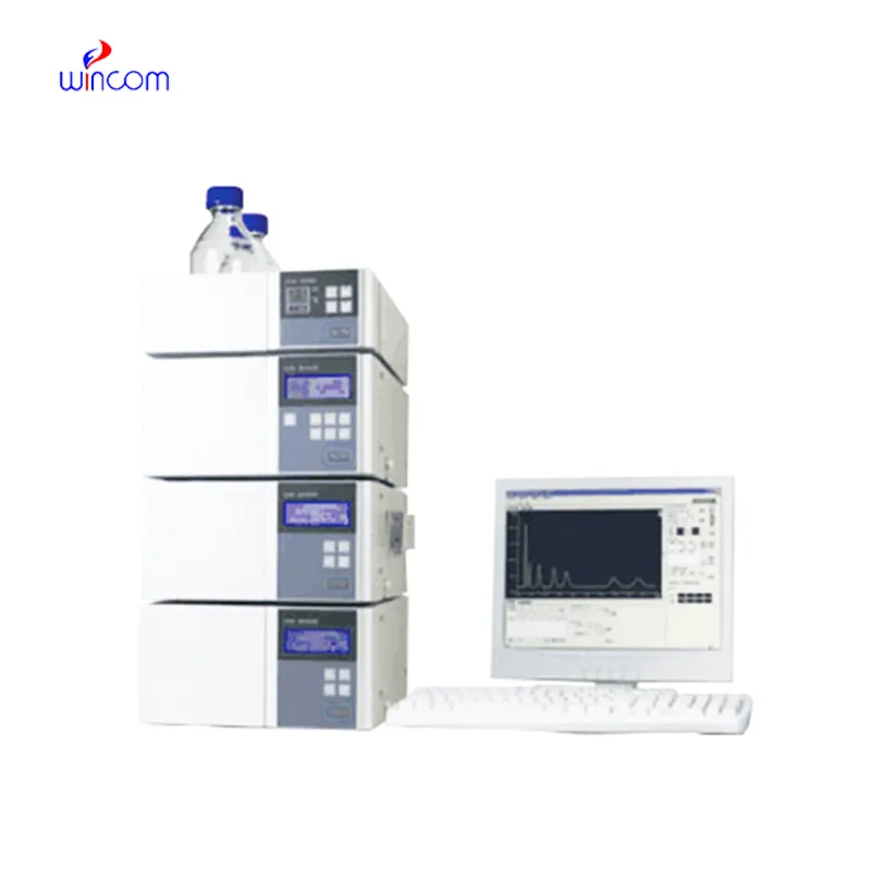
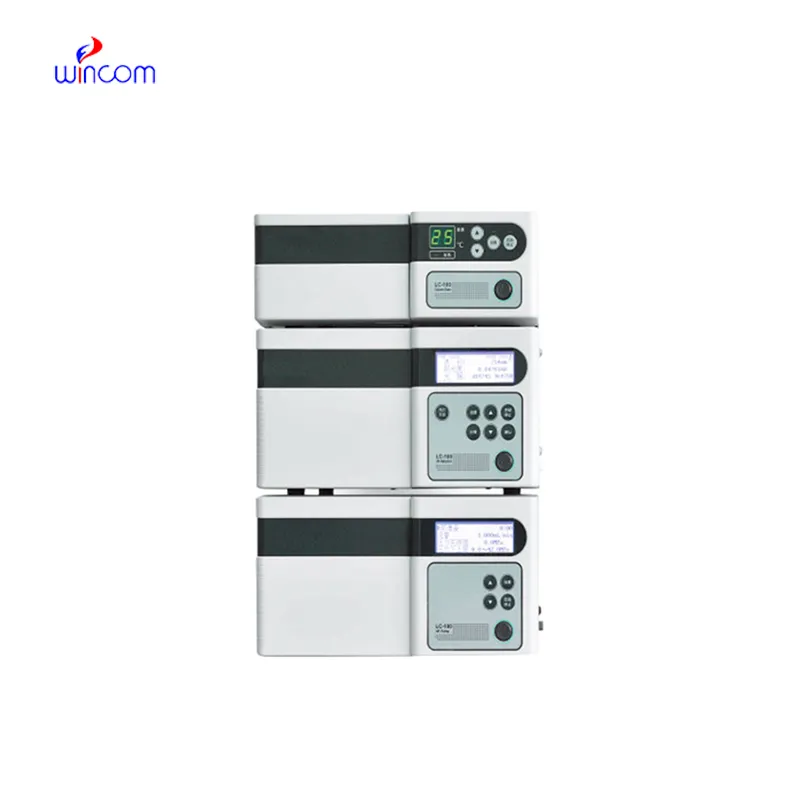
size exclusion hplc columns is commonly employed in laboratories to separate, identify, and quantify chemical compounds. The sample mixture is put through the columns along with the stationary phases and the different components interact with the stationary phase, thus the separation is done accurately. This process not only gives high resolution but also reproducibility thus it is a must-have tool for the research works in the area of drugs, pollution, and food control. Subsequently, when coupled with sensitive detectors, size exclusion hplc columns facilitates the precise measurement of minor concentrations. The method versatility produces so much that it has become a necessity in a routine analysis and complex research applications where it is positioned as an essential instrument in contemporary analytical chemistry and experimental workflows.
Q: What is HPLC used for in laboratories? A: HPLC turns out to be one of the most significant and essential analytical methods in laboratories equipped with the chemical compound analysis, separation, identification, and quantification of their presence in complex samples which are the research, clinical, and pharmaceutical applications. Q: How does HPLC separate compounds? A: The HPLC separation technique is based on the different affinities of the compounds to the stationary phase and mobile phase within the chromatography column. Q: Can HPLC analyze biological samples? A: Yes, it is certainly possible to carry out analyses on various biological fluids such as blood, serum, urine, etc. for the detection of metabolites, drugs, and biomarkers. Q: How often should HPLC columns be replaced? A: The replacement of the columns must be done according to the manufacturer instructions or when the performance begins to decline, which is quite usual after heavy use or contamination. Q: What detectors can be used with HPLC? A: The analysis type determines the use of, among others, UV, fluorescence, refractive index, and mass spectrometry detectors as the common detectors.
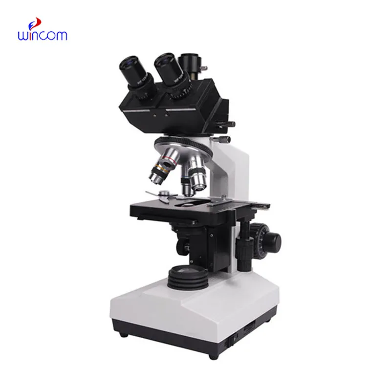
I’ve used several microscopes before, but this one stands out for its sturdy design and smooth magnification control.
This ultrasound scanner has truly improved our workflow. The image resolution and portability make it a great addition to our clinic.
To protect the privacy of our buyers, only public service email domains like Gmail, Yahoo, and MSN will be displayed. Additionally, only a limited portion of the inquiry content will be shown.
Hello, I’m interested in your water bath for laboratory applications. Can you confirm the temperat...
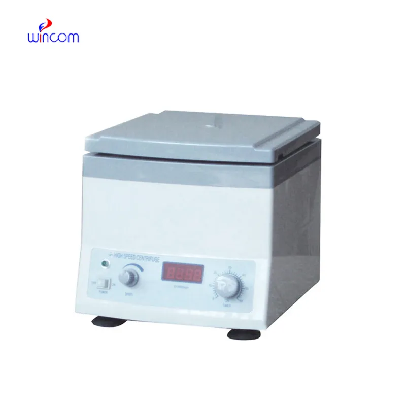
Hello, I’m interested in your centrifuge models for laboratory use. Could you please send me more ...
E-mail: [email protected]
Tel: +86-731-84176622
+86-731-84136655
Address: Rm.1507,Xinsancheng Plaza. No.58, Renmin Road(E),Changsha,Hunan,China

 af
af
 es
es
 ar
ar
 tr
tr
 sw
sw
 pt
pt
 th
th
 ur
ur
 bn
bn
 ne
ne
 vi
vi
 km
km
 lo
lo
 de
de
 ru
ru
 fi
fi
 nl
nl
 fa
fa
 fr
fr
 ko
ko